Table of Contents
A Revolutionary Tool in Breast Specimen Imaging for Patient Care
As a nurse, there are so many moments where we feel the weight of our responsibility to ensure the best possible care for our patients. One of the most significant advancements I’ve experienced in my nursing career has been working with the Mozart Machine. This revolutionary tool has transformed how we handle breast specimen imaging, and I can confidently say that it plays a crucial role in improving patient outcomes.
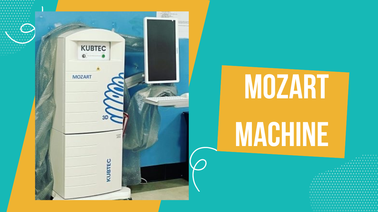
I remember when I was first introduced to the Mozart Machine. At that time, I had already been working in the operating room for a while, and I thought I had seen all the imaging tools that could help us in the fight against breast cancer. But then came this game-changer, and it immediately stood out because of its clarity and precision in producing 3D images of breast tissue.
In this blog post, I want to share with you how the Mozart Machine works and why it’s so vital. But more than that, I want to convey how using this technology has impacted me as a nurse, and how it has transformed the way we care for our patients.
The Mozart Machine: A Lifesaving Advancement in Imaging Technology
The first time I used the Mozart Machine, I knew we were stepping into a new era of breast specimen imaging. Before this, we relied heavily on traditional imaging techniques that, while useful, sometimes lacked the clarity needed for intricate breast tissue analysis. For women undergoing breast surgery—especially for something as daunting as breast cancer—the stakes are incredibly high. Getting the right diagnosis quickly can make all the difference in their treatment journey.
The Mozart Machine gives us a crystal-clear 3D image of breast tissue, helping to identify abnormalities like tumors that might not be as visible with older methods. As a nurse, I can’t emphasize enough how important this is. When I first saw those images come to life on the screen, I felt a mixture of awe and relief. It was a powerful feeling knowing that we could now catch problems much earlier, and that meant we were giving our patients a much better shot at beating the disease.
Step-by-Step Process of Using the Mozart Machine: From Setup to Review
Step 1: Preparation and Calibration – Setting the Stage for Success
Every time I prepare the Mozart Machine for imaging, I think about the importance of getting things just right. It’s not just about turning the machine on and pressing a button. There’s a process of calibration that ensures the images we capture are the best they can be. Imagine trying to take a picture with a camera that’s slightly out of focus—that’s what it’s like if the machine isn’t calibrated correctly. So, I take my time here.
I remember one particular morning when we had a patient scheduled for breast surgery. I was going through the calibration steps, adjusting the settings, and making sure everything was perfect. There was a bit of pressure in the air because we were all aware of the urgency of the situation—our patient had been diagnosed with a suspicious lump, and every minute felt precious. Once the machine was calibrated, I felt confident that we were ready to capture the best possible images for her.
Step 2: Entering Patient Information – Getting the Details Right
This is where attention to detail really matters. Before we even begin imaging, I make sure that all the patient’s information is entered correctly into the Mozart Machine’s system. It might seem like a small step, but it’s one of the most important. The images we capture are directly linked to the patient’s medical records, and any mistake here could cause confusion down the line. I collaborate closely with the radiology team and double-check everything to ensure accuracy.
I’ve had moments where I was rushing due to a busy schedule, and I’ve learned the hard way that it’s better to take a breath and slow down. The last thing anyone wants is to have mismatched data when dealing with something as serious as breast cancer. For me, this step isn’t just about data entry—it’s about making sure the patient’s story is told accurately through the images we capture.
Step 3: Capturing the Image – The Moment of Truth
Now comes the part that always gives me a sense of awe. After we’ve prepared everything, it’s time to actually take the image. I carefully place the breast specimen in the machine, ensuring it’s positioned perfectly. I’ve learned that even a slight shift in position can affect the quality of the image, so I pay close attention here.
Once the specimen is set, the Mozart Machine takes over. Within moments, it captures multiple high-resolution 3D images, providing us with a detailed view of the tissue. Watching those images pop up on the screen is always a profound moment for me. It’s like the machine is unveiling secrets hidden beneath the surface, allowing us to see things we wouldn’t have been able to spot with the naked eye.
One of the most emotional moments I’ve experienced was when we used the machine to capture images for a patient who had been fighting breast cancer for several years. This was a follow-up surgery, and we needed to ensure that the cancer had not returned. As the images appeared on the screen, I felt a lump in my throat. The clarity was astounding, and when the doctor confirmed that everything looked clear, I was overwhelmed with relief. I could see the same sense of peace wash over the patient when we gave her the news later that day.
Step 4: Reviewing the Data – A Critical Step for Nurses
After capturing the images, my job as a nurse continues. I transfer the images to our PACS system, where they are stored securely for the doctors and surgeons to review. But as a nurse, I don’t just walk away after this step. I take the time to review the images myself. We are often the first set of eyes on the data, and sometimes, we can spot potential issues or irregularities that may require further investigation.
I remember one case where, after reviewing the images, I noticed something that didn’t look quite right. I flagged it to the surgeon, and after a closer look, they agreed that further testing was needed. That moment was a reminder of how crucial it is for nurses to stay engaged throughout the entire process, from start to finish. We are an essential part of the care team, and our input can have a significant impact on patient outcomes.
Step 5: Collaborating with the Surgical Team – The Final Steps
The Mozart Machine is only one part of the process. Once the imaging is complete, I work closely with the surgical team to review the findings. We discuss the next steps based on the imaging results—whether that means additional surgery, further imaging, or other interventions.
In one memorable case, we had a patient whose imaging revealed a previously undetected abnormality. Because of the precision of the Mozart Machine, we were able to act quickly and adjust the surgical plan accordingly. It was a powerful reminder of how technology and teamwork come together to provide the best possible care for our patients.
The Human Side of Technology: Why the Mozart Machine Matters to Patients
For patients facing breast surgery, the emotional toll can be immense. Whether they are dealing with a cancer diagnosis or undergoing a biopsy for a suspicious lump, the uncertainty and fear can be overwhelming. I’ve had countless conversations with patients who express their anxiety about what lies ahead. It’s in these moments that I’m grateful for the clarity and precision that the Mozart Machine offers.
One patient, in particular, stands out in my memory. She was in her early 40s, with two young children at home, and had just been diagnosed with breast cancer. Understandably, she was terrified. After her surgery, I showed her the images we captured with the Mozart Machine and explained how this technology allowed us to get a clearer picture of her condition. Her relief was palpable. She said, “I feel like you’re able to see everything, like there’s nothing hidden, and that gives me hope.”
That’s why this machine matters so much—it gives patients hope. It gives them confidence that nothing is being overlooked, and that we are doing everything in our power to ensure their safety and well-being.
Looking to the Future: Embracing Technology in Nursing
As nurses, we are constantly evolving, learning, and adapting to new technologies. The Mozart Machine is just one example of how technology can revolutionize patient care. But it’s more than just the machine itself—it’s about how we use it, how we communicate with our patients, and how we integrate it into the overall care plan.
I believe that as healthcare continues to advance, nurses will play an even more critical role in harnessing these technologies. We are not just the operators of these machines—we are the caregivers, the advocates, and the ones who guide our patients through some of the most challenging moments of their lives.
External Resource:
For more information on advanced breast imaging technologies, visit this comprehensive guide tobreast imaging.
Internal Resource:
Check out my other blog post on Essential Steps for Specimen Management: A Guide for ScrubNurses.
Join our Email List Today!
We’d love to stay in touch and keep you updated with the latest insights and resources!
- Stay informed with exclusive content and updates.
- Receive expert tips and valuable resources directly in your inbox.
- Be the first to know about new articles, events, and more!
Fill out the form to subscribe now and be part of our growing community!
Let’s keep the learning and excitement going!
SUBSCRIBE TO MY YOUTUBE CHANNEL
If this post resonated with you, I also share calming visuals, quiet moments, and reflections on wellness over on my YouTube channel. You’re welcome to subscribe and join me there, whenever it feels right. Subscribe here/

Good day! I just want to give you a big thumbs up for your excellent information you have here on this post. I am coming back to your web site for more soon.